This blog will help healthcare professionals enhance the knowledge and skills necessary to navigate the complexities of airway management using Endotracheal (ET) Tubes. Managing a patient’s airway is a critical aspect of healthcare particularly in emergency and critical care settings. The proper use of endotracheal tubes is essential in ensuring the maintenance of a patient’s airway facilitating ventilation and providing oxygenation. These tubes are commonly used in various scenarios including surgeries intensive care units and pre-hospital care, to name a few.
This handbook serves as a valuable resource for both novice and experienced practitioners providing a wealth of information on the selection insertion and maintenance of endotracheal tubes. It delves into the anatomical and physiological considerations relevant to airway management, offering insights into the intricacies of patient assessment, tube sizing, and positioning.
Within this blog, you will find detailed descriptions of the different types of endotracheal tubes their respective indications and the associated procedural techniques. We explore the step-by-step process of safely and effectively inserting the tubes, highlighting the importance of proper positioning and securing to minimize complications and maximize patient outcomes.
Additionally, this handbook addresses the vital topic of troubleshooting and managing complications that may arise during the use of endotracheal tubes. It covers issues such as tube displacement, cuff leaks, and tube obstruction, offering practical solutions and strategies for quick intervention and resolution.
Whether you are a medical student a seasoned practitioner or anyone involved in the care of patients requiring airway intervention this handbook will serve as an indispensable companion in enhancing your skills and knowledge in this critical field. Prepare to delve into the world of endotracheal tubes and embark on a journey toward mastering airway management.
Understanding the Anatomy of Endotracheal (ET) Tubes
Endotracheal tubes (ET tubes) are indispensable tools in airway management, designed to establish and maintain a patent airway in patients requiring assisted ventilation or anesthesia. To fully comprehend the application and functioning of ET tubes it is essential to understand the anatomy of these devices and how they interact with the respiratory system.
ET tubes mainly consist of three main components which are the proximal end the main body and the distal end. The proximal end connects to the ventilation equipment and features an inflation port for the cuff. The main body is inserted through the mouth or nose and extends into the trachea. The distal end sits within the trachea and contains an inflatable cuff to create a seal. Understanding the anatomy of ET tubes is crucial for proper selection, insertion, and maintenance, ensuring effective ventilation and patient safety.
Understanding the anatomy of ET tubes is crucial for their proper selection, insertion, and maintenance. Factors such as tube size, cuff inflation, and positioning within the trachea significantly impact patient safety and ventilation effectiveness. It is worth noting that variations in ET tube design may exist based on specific clinical needs. Specialized tubes, such as double-lumen endotracheal tubes or tubes with additional ports, serve particular purposes in procedures such as lung isolation or bronchial hygiene.
By familiarizing oneself with the anatomy of ET tubes, healthcare professionals can optimize patient care by ensuring proper selection, correct placement, and appropriate maintenance of these vital airway management devices.
Types of Endotracheal (ET) Tubes
- Cuffed ET Tubes: Cuffed endotracheal tubes (ET tubes) are specialized airway management devices that feature an inflatable cuff near the distal end. The cuff serves a crucial role in providing a seal within the trachea, preventing the leakage of air and facilitating controlled ventilation. Cuffed ET tubes are commonly used in various clinical settings, including surgeries, intensive care units, and emergency medicine. They provide enhanced control over ventilation and help prevent complications associated with air leakage or aspiration. However, careful attention to cuff inflation, monitoring cuff pressure, and regular assessment of the patient’s airway is essential to ensure optimal airway management and patient safety.
- Uncuffed ET Tubes: Uncuffed endotracheal tubes (ET tubes) lack an inflatable cuff near the distal end. They allow for continuous airflow around the tube, potentially resulting in some air leakage. Uncuffed tubes are commonly used in specific situations, such as in pediatric patients or when cuff-related complications need to be minimized. Proper selection, sizing, and monitoring of uncuffed ET tubes are important for maintaining a patent airway and optimizing ventilation parameters. Alternative techniques may be considered for additional protection against aspiration. The use of uncuffed ET tubes depends on various factors, including patient characteristics, clinical indications, and individual clinician preferences. The decision to use an uncuffed tube should be made in consideration of the specific patient’s needs and the clinical setting. Proper selection, placement, and monitoring of uncuffed ET tubes are essential to ensure adequate ventilation and patient safety during airway management.
Endotracheal (ET) Tubes Size
Endotracheal tube size is a critical consideration when selecting and placing an ET tube. The appropriate tube size depends on factors such as the patient’s age gender and clinical indication. Some key points to understand about endotracheal tube sizing are:
- Internal Diameter: ET tube sizes are typically measured by their internal diameter in millimeters (mm). The internal diameter determines the amount of airflow and ventilation provided to the patient.
- Age-Based Recommendations: General guidelines exist for selecting the appropriate tube size based on the patient’s age. For pediatric patients, tube sizes decrease with younger age groups.
- Individual Variation: It is important to remember that individual patients may have unique anatomical characteristics that require deviations from age-based recommendations. Factors such as body size airway anatomy and underlying medical conditions should also be considered when selecting the appropriate tube size.
- Confirming Proper Fit: After inserting the ET tube, healthcare professionals should confirm its proper fit by assessing signs such as chest rise, breath sounds, and end-tidal carbon dioxide (ETCO2) waveform on capnography. These indicators help ensure adequate ventilation and appropriate tube sizing.
- Tube Positioning: Proper positioning of the ET tube is crucial for optimal ventilation and preventing complications. The tube tip should ideally be positioned above the carina (the point where the trachea branches into the bronchi) to minimize the risk of unilateral lung ventilation or accidental extubation.
- Special Considerations: In specific clinical scenarios, such as patients with restricted airways or those requiring lung isolation, specialized endotracheal tubes with unique features, such as double-lumen or bronchial blocker tubes, may be used. These specialized tubes allow for selective lung ventilation and enhanced procedural control.
The selection of an appropriate ET tube size is important for effective treatment and patient safety. Healthcare professionals should consider age-based recommendations and individual patient factors, and confirm proper fit through clinical assessment to ensure optimal ventilation and tube placement.
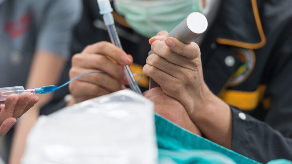
ET Tube Insertion/Endotracheal Intubation
Endotracheal intubation is a procedure that involves the insertion of an ET tube through the mouth or nose into the trachea to establish and secure a patient’s airway. It is a common intervention used in various healthcare settings including emergency medicine anesthesia and critical care. Some key points to understand:
- Purpose: The main goal of endotracheal intubation is to ensure a clear and secure airway allowing for mechanical ventilation or the administration of anesthesia when needed. It ensures a patent airway, allows for controlled ventilation, and protects the lungs from aspiration.
- Indications: Endotracheal intubation may be indicated in situations where a patient’s airway is compromised or anticipated to deteriorate. Examples include respiratory failure, severe trauma, coma, impending airway obstruction, or surgical procedures requiring general anesthesia.
- Procedure: Endotracheal intubation involves several key steps. These typically include pre-oxygenation of the patient, administration of appropriate sedation and muscle relaxation, visualization of the vocal cords using laryngoscopy, the passage of the ET tube through the vocal cords into the trachea, confirmation of proper tube placement, and securing the tube in place.
- Equipment: The equipment used for endotracheal intubation includes laryngoscopes (with blades for visualization), ET tubes of appropriate size, stylets (optional), syringes for cuff inflation, and devices for confirming tube placement (such as end-tidal carbon dioxide detectors).
- Complications: Endotracheal intubation is generally a safe procedure but it also carries potential risks and complications. These can include injury to the teeth, lips, tongue, vocal cords, or trachea; dislodgment or malpositioning of the tube; aspiration of gastric contents; and complications associated with the use of sedation or muscle relaxants.
- Verification of Tube Placement: Proper verification of tube placement is essential to ensure that the tube is positioned correctly within the trachea. This is typically done by assessing chest rise, breath sounds, and capnography readings (end-tidal carbon dioxide levels).
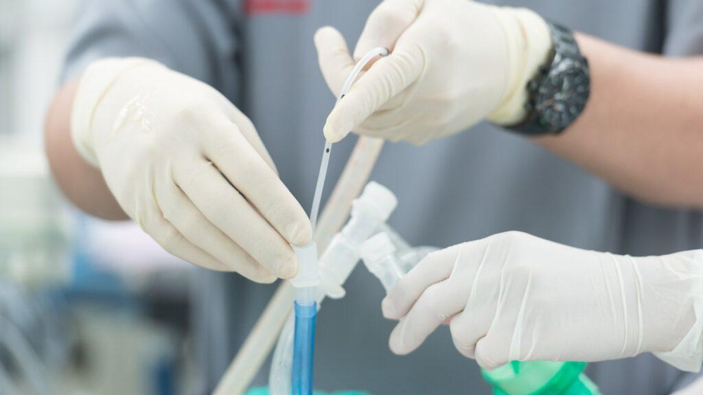
- Maintenance and Monitoring: Once the endotracheal tube is in place, it requires appropriate maintenance, including cuff pressure monitoring, regular suctioning, and ongoing assessment of ventilation parameters. Continuous monitoring of vital signs, oxygenation, and ventilation is crucial.
Endotracheal intubation is a complex procedure that requires skill, training, and careful attention to detail. It is typically performed by healthcare professionals with expertise in airway management such as anesthesiologists emergency physicians or critical care specialists. Proper technique, adherence to safety protocols, and vigilant monitoring are essential to ensure successful airway management and patient well-being.
ET Tube Removal/Endotracheal Extubation
ET tube removal also known as extubation is a procedure performed to remove the ET tube from a patient’s airway once it is no longer needed. Extubation is typically conducted when a patient’s condition has improved, and they can adequately maintain their own airway and breathe independently. Here are some key points to understand about ET tube removal:
- Assessment of Readiness: Before considering extubation, healthcare professionals assess the patient’s readiness by evaluating various factors, such as respiratory status, oxygenation, consciousness level, ability to protect the airway, and the underlying reason for intubation. Criteria for readiness may vary based on individual patient circumstances and clinical guidelines.
- Preparations: Adequate preparations are made before extubation to ensure a smooth and safe process. This may involve having appropriate personnel and equipment on standby, ensuring the patient is adequately oxygenated and stable, and confirming the availability of airway management alternatives if needed.
- Extubation Procedure: The actual extubation procedure involves the following steps:
Oxygenation: It helps to increase oxygen levels in the body.
Suctioning: The airway is suctioned to remove any secretions or debris.
Cuff Deflation: If the ET tube has an inflatable cuff, it is gradually deflated while assessing for air leakage and the patient’s tolerance.
Tube Removal: The ET tube is gently and smoothly removed from the patient’s airway during a coordinated effort, typically during a period of exhalation or under the guidance of a healthcare professional. - Post-Extubation Care: Following extubation the patient is closely monitored for any signs of respiratory distress airway obstruction or other complications. Vital signs, oxygen saturation, and respiratory effort are assessed regularly to ensure stability and adequate oxygenation.
- Airway Management Alternatives: In some cases after extubation patients may require alternative airway management methods such as a nasal cannula face mask or non-invasive positive pressure ventilation (NIPPV) to provide supplemental oxygen and support if needed.
- Documenting and Reporting: Accurate documentation of the extubation procedure, the patient’s response and any relevant observations are essential for continuity of care and communication among healthcare providers.
Extubation should be performed by experienced healthcare professionals who are knowledgeable about airway management and have the necessary skills to handle potential complications. Close monitoring and prompt intervention are crucial to managing any adverse events that may arise after extubation and to ensure patient safety and well-being.
ET Tube Complications
Common complications associated with endotracheal tubes (ET tubes) include:
- Trauma and injury to airway structures during insertion or removal.
- Tube misplaced into the esophagus instead of the trachea.
- Cuff-related complications, such as overinflation, underinflation, pressure injuries, or air leakage.
- Objective of gastric contents and oral secretions.
- Ventilator pneumonia due to bacterial colonization.
- Dislodgement or accidental extubation of the tube.
- Tracheal stenosis (narrowing) or ulceration from prolonged intubation.
- Psychological distress and discomfort for the patient.
Preventive measures, proper monitoring, and prompt management of complications are essential in ensuring patient safety and minimizing adverse events.
ET Tube Infections
ET tube infections also known as ventilator-associated pneumonia can occur in patients who are intubated and mechanically ventilated for a prolonged period. Key points about ET tube infections include:
- Risk factors: Prolonged intubation, prolonged mechanical ventilation, immunosuppression, and inadequate infection prevention measures increase the risk.
- Pathogens: Bacteria are common causes of infections along with fungi in certain cases.
- Mechanisms: Bacteria can enter the lower respiratory tract via the ET tube, adhering to its surface and forming biofilms.
- Symptoms: Fever, increased secretions, changes in sputum, and respiratory deterioration may indicate an ET tube infection, but further tests are necessary for a definitive diagnosis.
- Prevention: Hand hygiene, sterile insertion technique, oral care, elevation of the head of the bed, and minimizing intubation duration are important preventive measures.
- Diagnosis: Clinical assessment, imaging, laboratory tests, and sometimes bronchoscopy aid in diagnosing ET tube infections.
- Treatment: Antibiotics targeting identified pathogens are typically used, with prompt initiation to improve outcomes.
- Ventilator bundle: Implementing a ventilator bundle, including evidence-based practices, helps reduce ET tube infections by addressing various factors.
Preventing ET tube infections requires a comprehensive approach involving infection prevention, timely diagnosis, and appropriate management based on established guidelines.
Care and Maintenance of ET Tubes
To ensure proper function and minimize complications, here are key points for the care and maintenance of endotracheal tubes (ET tubes):
- Regularly assess tube placement, position, and stability.
- Monitor and maintain appropriate cuff inflation and pressure.
- Perform regular suctioning to clear respiratory secretions.
- Practice good oral hygiene to prevent infections.
- Prevent tube obstruction by promoting coughing, humidification, and hydration.
- Secure the tube properly to prevent accidental displacement.
- Monitor vital signs, oxygenation, and document assessments.
- Replace tubes periodically according to guidelines.
- Collaborate and communicate with the healthcare team.
Following these measures promotes the safe and effective management of ET tubes.
ET Tubes From Leading Indian Manufacturers
Aurolac
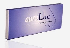
Aurolac, manufactured and supplied by Aurolab, is regarded as one of the best intubation sets available in India. With its numerous advantages, Aurolac is particularly well-suited for use in standard Dacryocystorhinostomy (DCR) surgeries. DCR is a technique that involves creating a shunt-like anastomosis between the lacrimal sac and the nasal mucosa to prevent nasolacrimal duct occlusion. The Aurolac lacrimal intubation set comprises two flexible stainless steel probes connected by a hollow medical-grade silicone tubing. This combination offers excellent biocompatibility and ensures a longer retention time. Aurolac facilitates easier insertion through the canaliculi and nasolacrimal duct, contributing to the success of the DCR procedure. The silicone tube, placed temporarily for a specified duration of six to twelve months, keeps the new tear drain open during the healing process.
Tracheostomy Recovery Kit
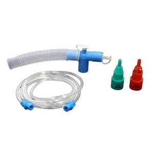 Tracheostomy Recovery Kit is a high-quality medical product manufactured by Nishi Medcare, designed to aid in the recovery process after tracheostomy surgery. This comprehensive kit provides all necessary tools and supplies for a smooth and efficient recovery.
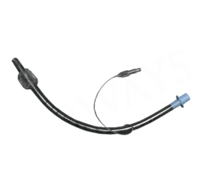
Airways Surgical Private Limited, India’s leading manufacturer and global supplier of respiratory care and critical care support products, presents the Airoline Endotracheal Tube. Made from non-toxic, transparent, thermosensitive silliconised polyvinyl chloride, this tube has undergone rigorous implant testing to ensure its safety and reliability. The Airoline Endotracheal Tube features a 15 mm connector for secure attachment to ventilation equipment. It is equipped with a radio-opaque line and a smooth tip, aiding in accurate placement. The tube incorporates 1 cm and 2 cm indicator marks, facilitating proper positioning relative to the vocal cords. A one-way valve pilot balloon labeled with the size allows quick identification and displays intra-cuff pressure. With a high-volume, low-pressure cuff and a Murphy eye, the Airoline Endotracheal Tube ensures efficient airway management for patients requiring intubation.
Endotracheal Tube
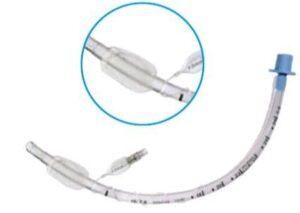
The Endotracheal Tube, manufactured by Aurus MedTech Pvt. Ltd., is a vital medical device employed for airway management and mechanical ventilation. Aurus MedTech Pvt. Ltd. is a reputable manufacturer known for its commitment to excellence. This tube is available in various sizes and types, offering versatility to healthcare professionals. Notable features of the Endotracheal Tube include a subglottic suction port, a Murphy eye for reduced trauma during intubation, and a Magill curve for ease of insertion. It is extensively used in general anesthesia, intensive care, and emergency medicine. To ensure optimal performance and patient safety, the proper placement of the tube must be verified, cuff pressure monitored, and regular suctioning and care provided to prevent complications. With its comprehensive features and meticulous design, the Endotracheal Tube from Aurus MedTech Pvt. Ltd. provides a reliable solution for airway management.
3055 – Tracheostomy Tube
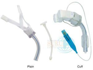
SURU International Pvt. Ltd., the leading manufacturer and global supplier of Endotracheal Tubes in India, presents the 3055 – Tracheostomy Tube. This specialized tube is designed for long-term tracheostomy needs. Manufactured from non-toxic implantation tested siliconised PVC, the 3055 – Tracheostomy Tube ensures the protection of delicate mucosal tissues. It is equipped with a soft flexible flange, allowing for easy fixation and minimizing patient discomfort. SURU International Pvt. Ltd. is dedicated to delivering high-quality tracheostomy tubes that meet the stringent standards of medical professionals worldwide.
Each of these medical products, including the Intubating Stylet, Aurolac, Airoline Endotracheal Tube, Endotracheal Tube, and 3055 – Tracheostomy Tube, offers unique features and benefits. They are manufactured by renowned Indian companies such as ONTEX Medical Devices Manufacturing Pvt. Ltd., Aurolab, Airways Surgical Private Limited, Aurus MedTech Pvt. Ltd., and SURU International Pvt. Ltd., ensuring exceptional quality and performance. With their advanced design and meticulous craftsmanship, these medical devices play a crucial role in airway management, respiratory care, and critical care support. Healthcare professionals can rely on these products to deliver safe and effective solutions for their patients’ medical needs.
Medzell: Revolutionizing Medical Device Promotion
In your journey to explore the world of endotracheal tubes it’s essential to stay updated with the latest advancements in the medical industry. Medzell, a futuristic B2B platform, is at the forefront of promoting Indian medical devices in emerging markets. With its innovative approach and vast network, Medzell connects manufacturers, distributors, and healthcare professionals, facilitating the exchange of knowledge and expertise. Stay informed about the latest developments in the medical device landscape with Medzell.
Conclusion
Endotracheal tubes play a vital role in airway management, ensuring the provision of adequate oxygenation and ventilation during medical procedures. From understanding their anatomy and selecting the right size to master intubation techniques and preventing complications, this blog has covered essential aspects of ET tubes. By expanding your knowledge and expertise in this field, you empower yourself to provide optimal patient care and enhance overall outcomes. Stay curious, stay informed, and continue exploring the remarkable world of endotracheal tubes.