In the realm of eye care, retinal cameras have emerged as indispensable tools for diagnosing and monitoring various ocular conditions. These advanced imaging devices capture high-resolution images of the retina, enabling healthcare professionals to detect and track changes in the eye. In this blog, we will delve into the world of retinal cameras, discussing their types, benefits, criteria for choosing the right one, how to use them effectively, maintenance tips, and the latest innovations in retinal camera technology. Join us as we explore the cutting-edge advancements in eye care with Medzell, your trusted B2B platform for medical supplies.
Types of Retinal Cameras
- Traditional Fundus Cameras: These are the most common type of retinal cameras, capturing detailed images of the retina using a flash or non-flash illumination. They are versatile and suitable for general retinal imaging.
- Handheld Retinal Cameras: Handheld retinal cameras offer portability and ease of use, making them ideal for remote or point-of-care settings. They are compact, lightweight, and often equipped with wireless connectivity for seamless data transfer.
- Widefield Retinal Cameras: Widefield retinal cameras provide a wider field of view, allowing for the capture of a larger portion of the retina in a single image. They are particularly useful for diagnosing and monitoring conditions that affect the peripheral retina.
Benefits of Retinal Cameras
- Early Detection of Eye Conditions: Retinal cameras enable healthcare professionals to detect early signs of eye conditions such as diabetic retinopathy, macular degeneration, and glaucoma. Early detection allows for timely intervention and better patient outcomes.
- Monitoring Disease Progression: By capturing high-resolution images of the retina over time, retinal cameras facilitate the monitoring of disease progression and treatment efficacy. This helps healthcare professionals make informed decisions regarding patient management.
- Patient Education and Engagement: Retinal images provide a visual representation of the patient’s eye health, allowing for better patient education and engagement. Visual evidence can help patients understand the importance of regular eye examinations and adherence to treatment plans.
Criteria for Choosing the Right Retinal Camera
- Image Quality: Look for a retinal camera that offers high-resolution imaging capabilities to ensure clear and detailed retinal images for accurate diagnosis and monitoring.
- Ease of Use: Consider the user-friendliness of the retinal camera, including features such as autofocus, intuitive controls, and ergonomic design. This ensures efficient and comfortable operation, especially during prolonged use.
- Connectivity and Integration: Check if the retinal camera offers seamless integration with electronic health record (EHR) systems or other imaging devices. This enables efficient data management and enhances workflow in healthcare settings.
How to Use Retinal Cameras Effectively
- Patient Preparation: Ensure that the patient’s pupils are dilated adequately for optimal retinal imaging. Follow the recommended guidelines for pupil dilation and provide clear instructions to the patient.
- Proper Alignment: Position the retinal camera correctly to align with the patient’s eye. Follow the manufacturer’s instructions for proper alignment and focus adjustment to obtain clear and accurate retinal images.
- Image Documentation: Capture multiple images of different areas of the retina to ensure comprehensive documentation. Pay attention to capturing the macula, optic disc, and peripheral retina, as per the patient’s condition and requirements.
Maintenance Tips for Retinal Cameras
- Cleaning and Disinfection: Regularly clean the camera’s lenses, eyepieces, and other surfaces using recommended cleaning solutions and techniques. Follow proper disinfection protocols to maintain a sterile environment.
- Calibration and Quality Assurance: Periodically calibrate the retinal camera to ensure accurate and consistent image capture. Implement quality assurance measures, such as regular image analysis and comparison, to monitor the camera’s performance.
- Software Updates: Stay updated with the latest software releases and firmware updates provided by the manufacturer. These updates often include bug fixes, performance enhancements, and new features that improve the camera’s functionality.
New Technologies and Innovations in Retinal Cameras
- Artificial Intelligence (AI) Integration: Retinal cameras are now incorporating AI algorithms to assist in the detection and analysis of retinal abnormalities. AI-powered image analysis can help healthcare professionals identify subtle changes and provide more accurate diagnoses.
- Optical Coherence Tomography (OCT) Integration: Some retinal cameras now offer integrated OCT technology, allowing for simultaneous retinal imaging and cross-sectional analysis. This provides a more comprehensive assessment of retinal health and aids in the diagnosis of complex conditions.
- Telemedicine Capabilities: Retinal cameras with telemedicine capabilities enable remote consultation and diagnosis, particularly useful in underserved areas or during emergencies. Real-time transmission of retinal images allows for timely expert opinions and reduces the need for patient travel.
Overall, these advancements in retinal camera technology have greatly improved the accuracy of diagnosis and enhanced the ability of healthcare professionals to provide exceptional eye care.
AI Algorithms in Retinal Cameras Improve the Accuracy of Diagnosis
AI algorithms in retinal cameras improve the accuracy of diagnosis by leveraging the power of machine learning and pattern recognition. Here’s how it works:
Early Detection: AI algorithms can analyze retinal images and identify subtle abnormalities or early signs of eye conditions that may not be easily detectable by human observers. By comparing the captured images with a vast database of known patterns and abnormalities, the AI algorithm can flag potential areas of concern for further examination.
Image Enhancement: AI algorithms can enhance the quality of retinal images by reducing noise, improving contrast, and sharpening details. This ensures that healthcare professionals have access to clear and high-resolution images, enabling them to make more accurate assessments.
Automated Analysis: AI algorithms can automatically analyze retinal images and provide quantitative measurements of various parameters, such as the thickness of retinal layers or the presence of specific lesions. This automated analysis saves time and reduces the risk of human error, allowing healthcare professionals to focus on interpreting the results and making informed diagnoses.
Decision Support: AI algorithms can provide decision support by offering suggestions or highlighting areas of concern based on the analysis of retinal images. This assists healthcare professionals in their decision-making process, helping them prioritize cases, recommend appropriate treatments, or refer patients to specialists when necessary.
Continuous Learning: AI algorithms can continuously learn and improve their performance over time. As more retinal images are analyzed and correlated with clinical outcomes, the algorithm can refine its diagnostic capabilities, leading to even higher accuracy in the future.
By harnessing the capabilities of AI algorithms, retinal cameras can assist healthcare professionals in detecting and diagnosing eye conditions with greater accuracy, ultimately improving patient outcomes and enhancing the overall quality of eye care.
Top Manufacturer of Retinal Camera
3nethra classic 6.4 Retinal Camera
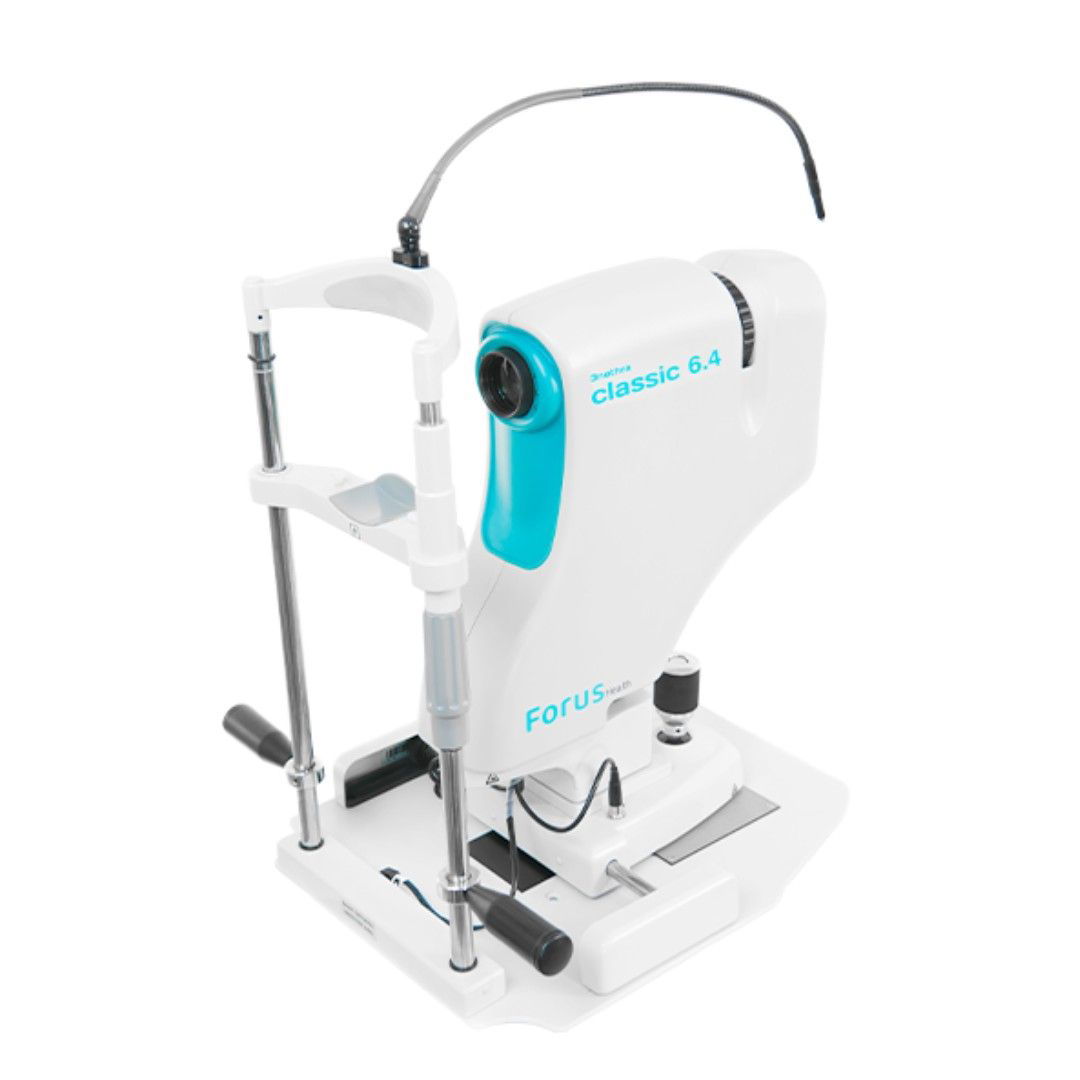
The 3nethra classic 6.4 Retinal Camera by Forus Health Pvt. Ltd. stands as a pinnacle in digital non-mydriatic fundus cameras, serving eye care professionals in South Africa, Nigeria, and Kenya. Boasting a high-resolution 6.4-megapixel sensor, this cutting-edge device enables the capture of detailed images of both anterior and posterior eye segments, facilitating comprehensive examinations for conditions like retinal diseases, glaucoma, cataracts, and corneal disorders. Widely esteemed in the specified regions, its top-tier image quality, and diagnostic prowess empower healthcare professionals with accurate information for informed decision-making, reinforcing its position as a reliable and affordable solution.
This retinal camera’s remarkable features include non-invasive scanning without pupil dilation, ensuring patient comfort, and detailed imaging for precise diagnosis and monitoring of various eye conditions. With a wide-angle view of the retina, it allows comprehensive examinations, aiding in early disease detection like diabetic retinopathy. The user-friendly interface, swift scanning process, and portability enhance accessibility and efficiency in diverse healthcare settings, including remote or mobile clinics. The 3nethra classic 6.4 emerges not just as a technological marvel but as a practical, indispensable tool in advancing eye care across South Africa, Nigeria, and Kenya.
Conclusion
Retinal cameras have revolutionized eye care by enabling early detection, monitoring, and patient engagement in the management of ocular conditions. By understanding the different types, benefits, criteria for choosing, proper usage, and maintenance tips, healthcare professionals can make informed decisions when selecting a retinal camera. Stay at the forefront of eye care innovation with Medzell, your trusted B2B platform for medical supplies. Embrace the power of retinal cameras and enhance your ability to provide exceptional eye care today!